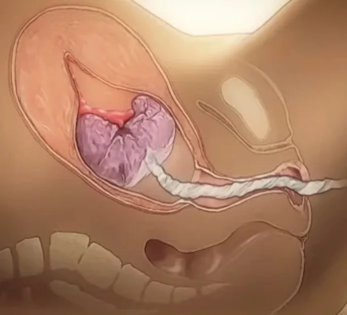
The Third Stage of Labour
The third stage of labour is the period from the birth of the fetus until the complete delivery of the placenta and fetal membranes.
Definition
- The third stage of labour is the period from the birth of the baby until the complete expulsion of the placenta and membranes from the uterus.
- "The third stage of labour begins after the delivery of the fetus and ends with the expulsion of the placenta and membranes."
Duration
- Usually 5–15 minutes
- Considered prolonged if it lasts more than 30 minutes
Events of Third Stage of Labour
The third stage of labour begins after the birth of the baby and ends with the expulsion of the placenta and membranes.
1. Placental Separation
After the baby is born, the uterus contracts and retracts strongly. This reduces the placental implantation site, causing the placenta to separate from the uterine wall.
Mechanism of Separation
A. Schultze Method (Central Separation)-Schultze = Shiny Surface First + Silent Bleeding
- Separation starts from the center of the placenta.
- Blood collects behind the placenta (retroplacental hematoma).
- Placenta comes out fetal surface first (shiny surface).
- Less visible bleeding before placental delivery.
B. Matthews Duncan Method (Marginal Separation)-Matthews Duncan = Maternal Surface First + More Visible Bleeding
- Separation starts from the placental margin.
- Blood escapes between the membranes and uterine wall.
- Placenta comes out maternal surface first (rough surface).
- More visible bleeding before placental delivery.
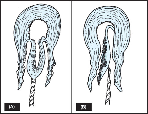
Schultze Method vs Matthews Duncan Method of Placental Separation
| Feature | Schultze Method (Central Separation | Matthews Duncan Method (Marginal Separation) |
| Site of separation | Starts from the center of the placenta | Starts from the margin (edge) of the placenta |
| Blood collection | Blood collects behind placenta forming retroplacental hematoma | Blood escapes between placenta and uterine wall |
| Visible bleeding | Less bleeding before placental delivery | More bleeding before placental delivery |
| Mechanism | Hematoma helps peel off the placenta from the center outward | Placenta separates progressively from one edge |
| Placenta delivered first | Fetal surface (shiny surface) appears first | Maternal surface (rough surface) appears first |
| Appearance during delivery | Placenta comes out like an inverted umbrella | Placenta slides out sideways |
| Membranes | Membranes usually follow neatly behind | Membranes may separate irregularly and tear |
| Frequency | More common (about 80%) | Less common (about 20%) |
| Blood loss | Comparatively less | Comparatively more |
2. Descent of the Placenta
After separation, the placenta descends from the upper uterine segment to the lower uterine segment and then into the vagina due to:
- Uterine contractions and retraction
- Maternal bearing-down efforts
- Gravity
3. Signs of Placental Separation
The following signs indicate that the placenta has separated:-
Maternal Signs
- Sudden gush of blood from the vagina
- Lengthening of the umbilical cord outside the vulva
- Uterus becomes firm, hard, and globular
- Uterus rises upward in the abdomen
4. Expulsion of Placenta and Membranes
The separated placenta is expelled through the birth canal by:
- Continued uterine contractions
- Maternal expulsive efforts
- Controlled cord traction (during active management)
- The membranes follow the placenta and are delivered intact.
5. Hemostasis (Control of Bleeding)
After placental delivery:
- Uterine muscle fibers contract and retract.
- Blood vessels at the placental site are compressed ("living ligatures").
- Blood clotting further seals the vessels.
- Postpartum hemorrhage is prevented
Management of Third Stage of Labour
Active Management of Third Stage of Labour (AMTSL)
Main Goal
- To ensure safe delivery of the placenta and prevent postpartum hemorrhage (PPH).
Objectives
- Promote effective uterine contraction and retraction.
- Facilitate early placental separation and expulsion.
- Reduce maternal blood loss.
- Prevent postpartum hemorrhage (PPH).
- Prevent uterine atony and retained placenta.
- Maintain maternal hemodynamic stability.
- Reduce maternal morbidity and mortality.
1. Preparation
- Wear sterile gloves.
- Keep emergency drugs and equipment ready.
- Ensure good lighting.
- Place a sterile tray for placental examination.
2. Administration of Uterotonic (Within 1 Minute After Birth)
- Oxytocin – Drug of Choice
- Oxytocin 10 IU IM or 5 IU slow IV
- Given after confirming no second baby is present.
- Misoprostol 600 µg orally is recommended as an alternative.
Important Note
- Methylergometrine/Ergometrine should be avoided in women with hypertension, pre-eclampsia, or heart disease.
- Oxytocin remains the first-line uterotonic for AMTSL.
Purpose:
- Promotes uterine contraction.
- Accelerates placental separation.
- Reduces blood loss.
- Prevents PPH.
3. Delayed Cord Clamping
Clamp and cut the cord after 1–3 minutes or when cord pulsations stop.
Benefits:
- Increases neonatal blood volume.
- Improves iron stores.
- Reduces neonatal anemia.
Note-Delayed cord clamping (1–3 minutes) allows transfer of 80–100 mL of placental blood and provides 40–50 mg/kg of iron.
4. Observe for Signs of Placental Separation
Look for:
- Sudden gush of blood.
- Lengthening of the umbilical cord.
- Uterus becomes firm and globular.
- Uterus rises in the abdomen.
5. Controlled Cord Traction (Brandt–Andrews Technique)
- Place one hand above the symphysis pubis to support the uterus (counter-traction).
- Hold the clamped cord with the other hand.
- Apply gentle downward traction during uterine contraction.
- Deliver the placenta slowly.

Purpose:
- Assists placental expulsion.
- Prevents uterine inversion.
- Reduces blood loss.
6. Delivery of Placenta and Membranes
- As the placenta appears at the vulva, hold it with both hands.
- Rotate it slowly to deliver membranes intact.
- Avoid pulling on membranes forcefully.
7. Examination of Placenta and Membranes
- Placenta
- Completeness of cotyledons.
- Maternal surface.
- Fetal surface.
- Membranes
- Complete amnion and chorion.
- No retained fragments.
- Umbilical Cord
- Length.
- Two arteries and one vein.
8. Uterine Massage-
- Immediately after placental delivery, palpate the fundus.
- Massage if uterus is soft.
Purpose:
- Maintains uterine contraction.
- Prevents uterine atony and PPH.
9. Assess Blood Loss and Maternal Condition
Monitor:
- Vaginal bleeding.
- Pulse.
- Blood pressure.
- Uterine tone.
- General condition of mother.
10. Perineal Examination
- Episiotomy wound.
- Perineal tears.
- Vaginal and cervical lacerations.
- Repair if necessary.
11. Documentation
- Time of placental delivery.
- Type of placental separation.
- Blood loss.
- Condition of placenta and membranes.
- Maternal vital signs.
- Drugs administered.